

Proc.Nati.Acad.Sci.USA Vol.76,No.4,pp.1858-1862,April 1979
secretory mutants/vesicles/membrane assembly
PETER NOVICK AND RANDY SCHEKMAN Department of Biochemistry,University of California,Berkeley,California94720 Communicated by Daniel E.Koshland,Jr.,January16,1979
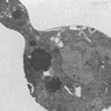
ABSTRACT Saccharomyces cerevisiae cells contain a small internal pool of the secretory enzymes invertase and acid phosphatase. This pool increases up to 8-fold at 37°C in a temperature-sensitive, secretion-defective mutant strain (sec 1-1). Cell division and incorporation of a sulfate permease activity stop abruptly at the restrictive temperature, while protein syn- thesis continues for several hours. Electron microscopy of mutant cells incubated at 37°C reveals a large increase in the number of intracellular membrane-bound vesicles, which are shown by histochemical staining to contain the accumulated acid phosphatase. The vesicles are removed and the accumulated enzymes are secreted when cells are returned to a permissive temperature in the presence or absence of cycloheximide. These results are consistent with a vesicle intermediate in the yeast secretory pathway and suggest that exocytosis maycontribute to cell-surface growth.
Protein secretion by plant and animal cells is mediated by a complex, highly organized series of membrane-bound structures (1, 2). The mechanism of glycoprotein secretion in Saccharomyces cerevisiae is less well understood. The tight coupling of protein synthesis with secretion and the low frequency of recognizable structures clearly associated with this process have prevented the formation of a coherent model. Membrane-bound vesicles have been implicated in the secretion of β-glucanases (3) and in bud (4) and division septum assembly (5); however, the role of vesicles in the secretion of acid phosphatase (6) and invertase (7) and in plasmalemma assembly has been less clear.
We have developed a genetic approach to the study of the secretory process in yeast. The analysis of a strain with a conditional, reversible block in the secretory pathway has allowed the identification of a vesicular intermediate in secretion and cell-surface growth.
MATERIALS AND METHODS
Materials. S. cerevtsiae haploid strain X2180-1A was from the yeast genetics stock center. A constitutive high acid phos- phatase-producing strain A137 (a, ACP 1-2, pho 80) and an acid phosphatase-defective strain Ela (a, acp 1) were obtained from P. Hansche (University of California, Davis; ref. 8). HMSF-1 (a, sec 1-1) was derived from X2180-1A. Standard genetic techniques were used to construct SF 150-5c (a,ACP 1-2,pho 80,sec 1-1), and SF 154-10A (a,acp 1,sec 1-1).
YPD medium contained 1% Bacto-Yeast Extract, 2% Bacto-Peptone, and 2% glucose. Wickerham's minimal medium (9) was used with the following modifications: for phosphatefree medium, potassium chloride replaced potassium phosphate; for sulfate-free medium, chloride salts replaced all sulfate salts. Unless otherwise indicated, the carbon source was 2% glucose. Petri plates contained minimal medium and 2% Difcoagar. Liquid cultures were grown in flasks with agitation, and the experiments were initiated with exponentially growing cells at an A600 of 1.5-2.5. When a change in the growth medium was required, the cells were collected by centrifugation, washed twice with distilled water, and resuspended in the new medium. The absorbance of cell suspensions was measured in a 1-cmquartz cuvette at 600 nm in a Zeiss PMQ II spectrophotometer; 1 A600 unit corresponds to 0.15 mg dry weight under all conditions of growth tested. Cell number was determined with a hemocytometer; buds were counted as cells.
Other reagents were obtained as indicated: Ethyl methanesulfonate, ρ-nitrophenylphosphate, glucose oxidase, ο-dianisidine, peroxidase, cycloheximide, and homocysteine thiolactone-HCI were from Sigma; H235SO4, L-[4,5-3H]leucine, L-leucine, and L-methionine were from ICN; mycostatin (nystatin) was from Calbiochem; glusulase was from Endo Laboratories (Garden City, NJ). Lyticase is a yeast lytic enzyme preparation (unpublished observations), useful in spheroplast formation (10). Fraction II (30,000 units/mg; 1 unit will lyse 0.2 A600 of logarithmic phase S. cerevisiae in 30 min at 30°C) was used.
Isolation of Secretory (sec) Mutants. X2180-1A cells were grown in YPD medium and treated with 3% ethyl methanesulfonate for 60 min at 25°C; the survival rate was 50-70%. The mutagenized culture was diluted with an equal volume of 12% sodium thiosulfate, and the cells were centrifuged and washed twice with distilled water. The cells were then grown in YPD medium for 8 hr at 24°C, and diluted aliquots were spread on minimal medium agar plates. After 3 days at 22°-24°C, 1600 colonies were replica-plated onto YPD medium and incubated overnight at 37°C. The temperature-sensitive clones (87/1600) were replica-plated onto phosphate-free minimal medium to derepress the synthesis of acid phosphatase, and after 10 hr at 24° or 37°C the replicas were stained for secreted acid phosphatase (8). The clones that showed temperature-sensitive secretion of phosphatase were screened for conditional secretion of invertase. Cultures grown at 24°C in liquid minimal medium containing 5% glucose were shifted to fresh medium containing 2% sucrose, and after 5 hr at 24° or 37°C the cells were centrifuged, washed with distilled water, and assayed for secreted invertase. Two clones showed conditional secretion but normal incorporation of 35SO42- into protein at 37°C (data not shown). The mutant loci designated sec 1-1 and sec 2-1 are nonallelic and recessive. Only sec 1-1 will be described in this report.
Assays. External (cell wall-bound) invertase was assayed at 37°C as described by Goldstein and Lampen (11); units of activity are μmol of glucose released per min. External acid phosphatase was assayed at 37°C as described by van Rijn etal. (12); units of activity are nmol of ρ-nitrophenol released per min. Sulfate permease activity was assayed at 37°C in 50 μM (NH4)2SO4 as described by Breton and Surdin-Kerjan (13); units of activity are nmol of SO42- uptake per min. Internal acid phosphatase and invertase were assayed in spheroplast lysates. Washed cells (10 A600 units) were resuspended in 2 ml of 1.4 M sorbitol/0.1 M potassium phosphate, pH 7.5/0.5 mM sodium azide/20 mM 2-mercaptoethanol/80 units of lyticase. After 45 min at 37°C the spheroplasts were centrifuged at 10,000 X g for 5 min, and the pellets were resuspended in 0.5 ml of 1% Triton X-100. Residual 2-mercaptoethanol present in the spheroplast lysate was eliminated with N-ethylmaleimide (0.4 mM), which was added after the first stage of the internal invertase assay. Protein synthesis was measured in 1-ml aliquots of minimal medium containing 1 A600 unit of cells and 50 nCi of L-[3H]leucine (0.33 Ci/mol, 1 Ci = 3.70 X 10¹⁰ Bq). Incorporation was stopped after 10 min with 1 ml of cold trichloroacetic acid (20%). After 1 hr at 0°C the mixtures were filtered on Whatman GF/A filters; the filters were washed and dried, and the radioactivity was measured in a Searle Delta 300 liquid scintillation counter.


